
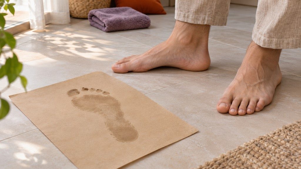
Foot arches can look different when you sit and stand. A wet footprint shows sole contact. A clinician considers the appearance together with pain, flexibility and walking.
A flat feet test at home can start by comparing how the arch on the inside of your foot looks while you sit and stand. A wet footprint can also show which parts of the sole touch the surface. During a clinical assessment, these observations are considered together with pain, flexibility and the way you walk. [1, 2, 3]
Look while seated. Sit barefoot and note the shape of the arch on the inner edge of each foot while it is free of your body weight. Try to compare both feet from a similar angle.
Compare while standing. If you can stand comfortably and safely, place both feet on a dry, level surface and let your weight rest naturally on them. The arch may look lower in standing and more visible while seated. Note whether one foot looks different from the other and how long you have noticed the change. [1, 2]
Notice what happens when a child moves. If a child comfortably rises onto the toes of both feet on their own, the arch may become visible again. This can be consistent with flexible flat feet. Avoid prompting the movement when there is pain, a recent injury or difficulty balancing. A single-leg heel rise is a separate clinical assessment that a clinician may use when appropriate. [1, 4]
Lightly dampen the sole. Put a piece of paper on a flat, dry surface and make sure it cannot slip. If you can step safely, stand naturally on the paper for a moment and look at the print. Dry your foot afterwards. If balance is difficult, skip the print and use only the observations you can make safely.
The width of the print between the heel and forefoot shows how much of the middle of the sole contacted the paper. A broader contact area can be consistent with a low arch. Flexibility, the cause of pain and the distribution of pressure call for other forms of assessment. Even standardised footprint indices used in research vary in how accurately they screen for arch shape. Share the print as an observation if helpful and discuss any assessment or support needs with a clinician. [3, 5]
| Observation or assessment | Question it helps answer |
|---|---|
| Looking at the arch while sitting and standing | How does the arch change when it takes weight? |
| Wet footprint | Which parts of the sole touch the paper? |
| Clinical examination of movement and flexibility | How readily does the foot move between positions? |
| Walking observation and pressure measurement if indicated | How do movement and loading change during a step? |
| Weight-bearing X-ray if indicated | How are the bones aligned when standing? |
The clinician may ask when the change began and how it affects daily activities. Describe the location of pain, what brings it on and whether it limits walking. The clinician may compare the feet with and without weight on them, then examine heel alignment, joint movement and tenderness and observe walking. A heel-rise assessment may help the clinician examine movement and function when indicated. [2, 4]
Painless flexible flat feet are common in children. The arch can appear lower while standing and reappear when weight is taken off the foot. When the arch remains low with the foot unloaded and the foot seems stiff, an examination can clarify flexibility. Report any effect on everyday walking and play. [1, 6]
In adults, a newly developing change warrants medical assessment, particularly when it affects only one foot. Pain or swelling on the inner side of the ankle can accompany some tendon problems. Explain when the symptoms began and how they affect walking. [3, 4]
Foot pressure measurement shows how pressure is distributed across the sole under the conditions recorded, which may include standing or walking. The clinician interprets that information alongside symptoms and examination findings. X-rays show bone alignment. Routine imaging is generally unnecessary for typical painless, flexible flat feet in children. When pain, stiffness or a newly developing change calls for imaging, the clinician chooses the method that addresses the clinical question. [4, 6]
For more about what is measured and how the results are discussed, see the gait analysis guide. For children, the guide to flat feet in children explains how age, symptoms and support needs are considered.
Seek an assessment for recurring pain, stiffness, weakness, numbness or difficulty walking or balancing. Mention any new change or clear difference between the feet. Before the visit, write down when symptoms started and what activities or shoes make them more noticeable. If a new injury leaves you unable to bear weight, seek prompt medical assessment and leave the home checks for another time. [3, 7]
Home observations can help you describe how the arch changes with weight and whether one foot looks different. The cause and any need for support are assessed clinically. [2, 3]
It shows that a wider area of the midfoot touched the paper. This can be consistent with a low arch. Clinical assessment also considers flexibility, symptoms and walking. [5, 6]
This can be a sign of a flexible foot. Pain or difficulty moving still warrants assessment. [1, 6]
Painless flexible flat feet are common. When a child walks and plays comfortably, the appearance of a low arch alone generally requires no treatment. [1, 6]
Arrange medical assessment. Describe when the change began, any pain or swelling and whether walking has changed. [3, 4]
The clinician decides after examining the foot. Routine X-rays are generally unnecessary for typical painless, flexible flat feet in children. [4, 6]
A footprint records the shape of the area in contact with the surface. A pressure map shows how pressure is distributed under the recorded conditions. [5]
A recommendation depends on the examination, symptoms and intended use. If support is suggested, ask what it is intended to help with and how the benefit will be assessed. [1, 2]